
More than a year ago, UC Davis became the first veterinary hospital in the world to offer equine PET scans. Their veterinarians are continuing to see successes in their discoveries of the diagnostic capabilities of PET. Recently, a chronic lameness in an American Quarter Horse named Bella was finally solved due to the use of PET.
Bella, a 16-year-old American Quarter Horse mare, has historically suffered from chronic hind limb issues. She was previously diagnosed with bilateral osteoarthritis of the lower tarsal joints (hock), but intra-articular medication had failed to significantly improve her lameness on the right hind. Bella had been ridden by two small children for the past three years and is an integral part of their family. Due to the severity of this lameness, however, she could no longer be ridden. Committed to improving Bella’s condition, her family brought her to the UC Davis veterinary hospital.
Upon being presented to the Equine Surgery and Lameness Service, Dr. Larry Galuppo and his team performed an exhaustive lameness exam with diagnostic analgesia and used a computerized lameness locator system to confirm that the area causing her pain and lameness was the right hock. A possible surgical intervention consisting of fusing some of Bella’s tarsal joints was considered, but additional information to determine the exact location of the lesion and plan for the surgery was needed.
To determine this, Bella was enrolled in a clinical trial offering advanced imaging techniques unique to the UC Davis veterinary hospital. More than a year ago, UC Davis became the first veterinary hospital in the world to offer Positron Emission Tomography (PET) for its equine patients. Combined with Computed Tomography (CT), this state-of-the-art imaging process can pinpoint locations of lameness with more accuracy than CT alone or other forms of imaging.
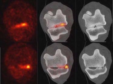
PET (left), fused PET/CT (center) and CT (right) images of Bella, showing the active bone changes (orange) responsible for Bella’s pain and lameness.
© UC Davis Veterinary Medicine
A team of veterinarians and technicians from the Anesthesia/Critical Patient Care and the Diagnostic Imaging services assisted Bella’s equine veterinarians to perform a PET/CT scan on her under general anesthesia. The PET/CT revealed that the main cause of pain in Bella’s hock was osteoarthritis of the talocalcaneal joint, rather than the degenerative changes in her distal intertarsal and tarsometatarsal joints identified with radiographs and MRI. This joint is rarely affected in horses, and its location and configuration make diagnostic and therapeutic procedures quite challenging. The PET/CT findings explained why the previous treatment had been unsuccessful and suggested that surgical intervention to the distal tarsal joints was not needed. While Bella was still under general anesthesia, a CT guided injection of the talocalcaneal joint was performed. Bella recovered well from the anesthesia following the procedure.
Four weeks after the PET/CT and the CT guided injection, Bella’s condition significantly improved, and she could be ridden again. The owners were completely satisfied with the therapeutic and diagnostic procedures performed, and were thrilled to have Bella returned to a healthy state.
For Bella, PET/CT was an absolute game changer since it allowed precise identification and a targeted treatment towards the affected joint.
UC Davis veterinarians, led by Drs. Mathieu Spriet and Pablo Espinosa, are continuing to see successes in their discoveries of the diagnostic capabilities of PET through this clinical trial and other research focused on advancing the clinical applications of this new modality.
Source: UC Davis Veterinary Medicine press release
READ THE LATEST NEWS ARTICLES HERE

https://www.equestrianlife.com.au/articles/US-university-uses-PET-and-CT-scanning-to-diagnose-lameness