|
This article has appeared previously with Equestrian Life. To see what's in our latest digital issue, click here.
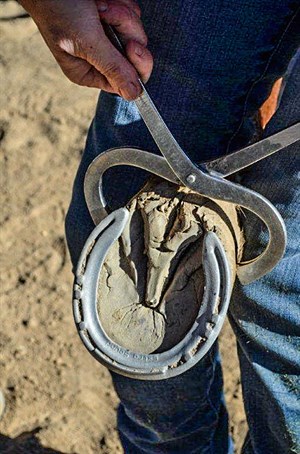
Hoof testers can be a useful tool.
© Maxine Brain
Zooming in on lameness
I’m talk lameness this issue, rather than soundness as many people ask, because soundness encompasses many aspects of equine health and behaviour, apart from just lameness.
BY MAXINE BRAIN
EVERYONE KNOWS WHAT a lame horse is, yet everyone’s perception may differ greatly. By definition, lameness is a “weakness or partial function of a leg, so that the gait is abnormal” (Blakiston’s Pocket Medical Dictionary). Essentially this means there is something wrong with either the structure or the functioning of a limb. Lameness can be the result of pain or mechanical dysfunction in one or more limbs, or can involve issues relating to the neck and spinal cord.
There are numerous causes of lameness which can be categorised into:
• Traumatic (bruises, torn muscles, tendon injuries and fractures).
• Degenerative (arthritis, ringbone).
• Infectious (cellulitis, foot abscesses).
• Congenital (club feet, osteochondritis, bone cysts).
• Metabolic (“tying up”); neurological (stringhalt, wobblers).
• Neoplastic (bone tumours) in origin.
In most cases I see, the causes are generally traumatic, infectious or degenerative in nature.
Before diagnosing the cause of lameness, it is important to categorise the type of lameness and grade its severity. This enables the vet to envisage possible causes, gauge the seriousness of the problem and record an accurate assessment for future reference.
Methods of classification can include whether the lameness affects the swing phase of the leg (when the leg is moving) versus the supporting limb (when the foot lands or the horse is weight-bearing on the limb), or a mixture of the two. Characteristics of the stride, the foot flight and the way the foot lands are also important factors to note. A common way to describe lameness is to grade the severity of it on a numeric scale. The following is a basic outline of a lameness score chart developed by veterinarians.
GRADE OBSERVATION
0 The horse is not lame at all, no gait anomalies present.
1 There is a mild lameness, not always apparent to the untrained eye.
2 There is an obvious lameness with a mild head bob or pelvic hike. It is not visible at the walk.
3 The lameness is easily seen with a more pronounced head bob or pelvic limb hike evident. (Grade 3 hind limb lameness often will produce a head bob as well as a pelvic hike, resulting in confusion of which leg is the lame one).
4 This is a severe lameness, often visible at the walk. When trotted it results in an exaggerated or extreme head nod and pelvic hike.
5 This is a non-weight-bearing lameness, so that the horse will physically carry the leg when asked to move forward.
Assessing lameness and diagnosing the cause can be simple sometimes and incredibly difficult at other times. A good history with a thorough clinical examination are essential at the outset. The use of imaging equipment should be reserved until the vet has made a clinical judgment on where the site of the lameness is and what the cause might be.
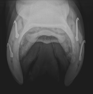
This is an x-ray of a navicular bone looking for evidence of “navicular disease”.
© Maxine Brain
Types of Diagnosis – what is right for your horse?
History: This helps categorise the lameness into a specific cause. A horse that goes out to work and suddenly pulls up lame is more likely to have a traumatic injury than, say, an infectious or neoplastic injury. A horse that has competed for years is less likely to have a congenital issue than a young horse competing for the first time.
Clinical Examination: The horse should be observed at rest then at a walk or a trot. Once the lame leg (or legs) is identified, it should be closely palpated and passively flexed to look for areas of swelling, heat or soreness. Hoof testers should be applied to the foot if no obvious cause is identified beforehand. Tendons should be palpated for signs of swelling and pain. Often it is good to compare responses to palpation on the good leg to ascertain if any pain is specific to that region. If a metabolic condition is suspected then a blood test looking at certain muscle enzymes (CPK, AST) should be done.
Flexion Tests: These are a very useful even though they have been bad-mouthed by many owners. Basically, the limb is flexed in a particular way, depending on what joint the vet wants to test, and the horse is made to trot off when the limb is released. The aim is to exaggerate the lameness to localise the specific area of soreness. For example, if a fetlock joint is flexed and is the site of the soreness, the horse will often trot off sorer. How long the limb is flexed for and how hard will vary depending on the person conducting the test. Some people believe these tests they can make a “sound” horse lame, but this is not a problem with the tests, it is a problem with their interpretation. Provided the tests are conducted in a consistent manner by an experienced person, they can result in a lot of useful information.
Nerve Blocks: Local anaesthesia is used to deaden or reduce sensation to specific areas of the leg. These can be extremely valuable for certain areas, particularly the lower limbs and joints. An improvement of around 60 per cent in lameness is considered positive and warrants further investigation of the region or joint that was blocked. I do not like to nerve-block horses that I am concerned may have a fracture, as there is a risk of further injury if the horse is allowed to use the limb fully when numbed.
Radiology: X-rays are very useful to examine bones but are of limited use for soft-tissue injuries. Due to the size of horses, there are limitations to what can be X-rayed with portable machines. Good quality X-rays of the limbs up to the elbow on the forelimb and stifle on the hind limb can be obtained on most digital machines, but shoulders require a skilled operator. X-rays of the pelvis and along the spine, with the exception of the neck and tail, require large fixed machines which are generally only in referral hospitals.
Ultrasonography: The ultrasound machine is a valuable diagnostic tool for soft-tissue injuries, particularly for examining tendons and ligaments. It can also be used for assessing fluid in joints and tendon sheaths; muscle tears; for examining the surface of bone; and to diagnose ailments such as a fractured pelvis or scapula (shoulder blade) which are not able to be radiographed.
Scintigraphy: A form of nuclear medicine where a radioactive isotope is injected into the bloodstream and concentrated into areas of damaged bone (and some soft tissue if visualised quickly). With the use of a gamma camera these “hot spots” are identified and assessed to find the site of lameness. The procedure is particularly valuable for diagnosing “stress fractures”, especially in the upper limbs and pelvis. These can be difficult to detect on a clinical examination and may not be visible on X-rays in the early stages. The other major use for scintigraphy is assessing poor performance when low-grade lameness is present in one or more limbs or the spine is involved. The procedure is normally only conducted at referral centres and is relatively expensive ($1500-$2500).
CAT (computed tomography): This is an advanced form of radiology which allows for more detailed images of bone. Multiple 2D images are collected and a 3D image is constructed so the clinician can obtain a detailed structure of the part of the limb being examined.
MRI (magnetic resonance imaging): This uses strong magnetic fields to image soft tissue and bone and gives a more detailed image than radiology or ultrasonography. The disadvantage is that MRI is limited mainly to the lower legs (knees or hocks and below).
There are basically two types available: a high-definition model which requires the horse to be anaesthetised; and a low-definition model which can be done on the standing horse. There are pros and cons with both machines. The HD machines are expensive to buy and require a large dedicated area; there are only a few in Australia. They produce a superior image but require a general anaesthesia for the horse. Whilst many horses undergo anaesthesia safely, there is a risk involved with any anaesthesia and the cost adds to the price of the MRI.
Low-definition machines can be conducted on the standing horse, but the horse still requires heavy sedation to stand still. The anatomical site of the lameness should be identified before an MRI scan, because the areas imaged are typically small and detailed. (About $2000 plus the cost of anaesthesia).
Once the site of lameness is identified and the cause established, the next step is to treat the lameness and prognosticate on the future of the horse... another topic worthy of its own article!
READ THE LATEST NEWS ARTICLES HERE

|