There are few diseases crueller to a horse than acute laminitis caused by toxins in the blood, or endotoxaemia. To make these horses even a little more comfortable during these episodes is a challenge; to save them is a feat.
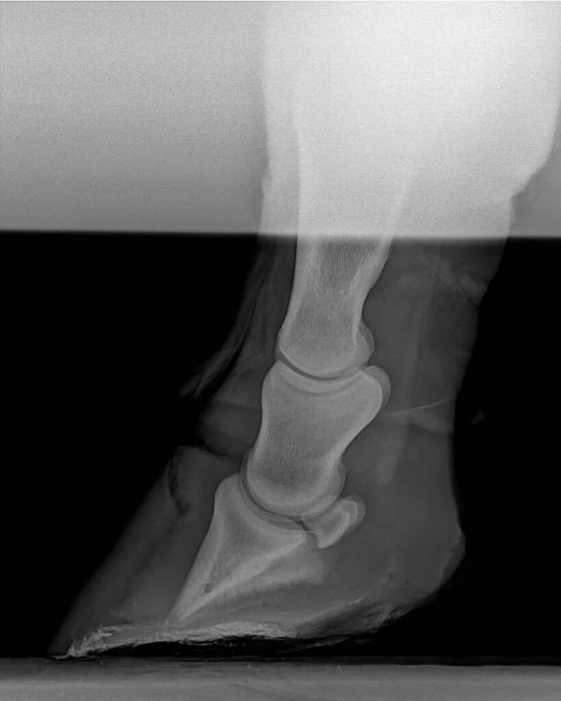
This x-ray shows a horse with severe laminitis where the pedal bone is bordering on penetrating down through the sole. The dark area between the hoof wall and the pedal bone is where there has been separation between the hoof wall and the pedal bone. Image supplied by Dr Maxine Brain.
Unlike the typical metabolic-induced laminitis which can occur slowly and is often mild to moderately painful during the initial stages, endotoxic-induced laminitis can occur within hours to days of an endotoxaemia event and render the horse almost completely immobile “overnight” due to the pain in the feet. This is where urgent veterinary attention is called for.
Laminitis is the inflammation of the lamellae in the feet of horses. Lamaelle are the finger-like epidermal and dermal projections incorporating blood vessels that intertwine and connect the hoof wall to the pedal bone. When the epidermal or dermal projections are inflamed, blood vessels are compromised and blood flow through them becomes markedly decreased or non-existent. The connection between the hoof wall and the pedal bone then breaks down, allowing the pedal bone to move within the hoof capsule.
There are a few different types of laminitis, and these are categorised based on the inciting cause. The more commonly seen laminitis is endocrinopathy laminitis and is the one we typically see in ponies and obese horses and is triggered by hyperinsulinaemia (high levels of insulin in the blood). This has been associated with horses/ponies that have hormonal conditions like Cushing’s disease or Equine Metabolic Syndrome.
The second type of laminitis is supporting limb laminitis and is induced when one limb is severely incapacitated and basically non-weight-bearing. With this type the horse places most of its weight onto the normal limb, which compromises the blood flow and induces laminitis in the “sound” limb.
The third type of laminitis and the topic of this article is septic, or endotoxaemia-induced, laminitis.
There are two main ways the pedal bone will move, with the more commonly known being rotation of the pedal bone. This is where the tip of the pedal bone moves downwards so there is a loss of the parallel orientation of the front of the pedal bone relative to the hoof wall. It is thought the deep digital flexor tendon that attaches to the back of the pedal bone pulls the back of the pedal bone up and then the tip of pedal bone, having no support from the damaged lamella, moves downwards. In severe cases the tip may penetrate the sole of the foot.
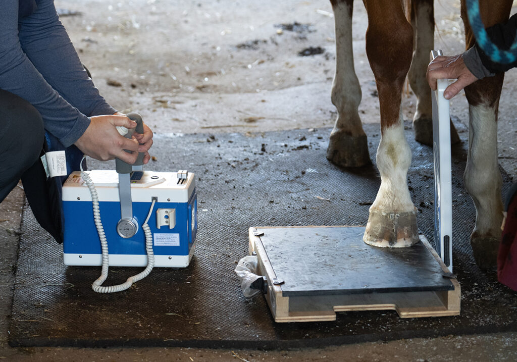
X-raying the feet will give an indication of the damage present.
A better way to determine the severity of damage is to do what is called a venogram on the feet. A tourniquet is placed around the lower limb and contrast medium injected into one of the main blood vessels that supplies the foot, and an X-ray is taken immediately following this injection. Contrast medium allows the blood vessels that have blood flowing through them to be seen on X-ray. Blood vessels that are no longer capable of allowing blood to flow through them remain black on the X-ray.
“X-raying the feet will
give an indication of
the damage present.”
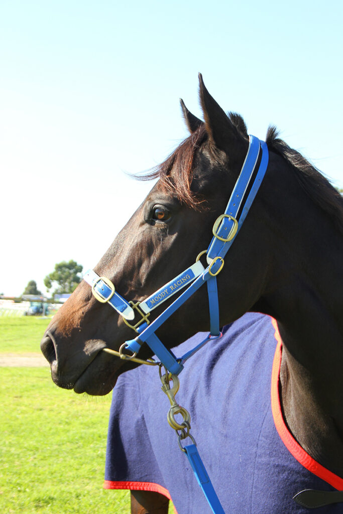
Champion racehorse Black Caviar was sadly euthanised following a bout of laminitis. It was reported the onset of laminitis was following a milk infection; therefore it’s plausible this was a case of acute laminitis caused by endotoxaemia and then exacerbated by complications associated with pregnancy. Image by Equestrian Life.
BETTER-INFORMED PROGNOSIS
By seeing the degree of damaged vessels, the veterinarian can give a better-informed prognosis and allow them to identify horses that have insufficient blood to the foot in a timely manner to be able to recommend euthanasia in horses with a hopeless prognosis.
Endotoxins are toxins produced within the body and originate from the cell wall of gram-negative bacteria that have acquired entry into the body. Once these toxins enter the bloodstream in high levels, they establish an endotoxaemia (“toxins in the blood”). The term endotoxaemia is also used synonymously to describe the clinical presentations seen when the endotoxins react with the body’s immune system, sending it into overdrive.
Endotoxins induce an inflammatory cascade that produces severe and often fatal responses. This is related to the uncontrolled release of small peptides that would in normal circumstances only be released in small quantities to facilitate an appropriate response to an inflammatory stimulus. Instead, certain cell types in the blood become over-reactive and secrete large quantities of cytokines that cause vascular system (blood) dysfunction, and this can include forming small blood thromboses (blood clots) that interfere with blood flow to the major body organs. This loss of oxygen flow to the tissues causes further cell death and can lead to major organ failure in septic horses and foals.
Whilst there is a well-established connection between endotoxic horses and the onset of laminitis following illness, the exact mechanism that causes the laminitis has not clearly been established. It may be as simple as the blood flow to the foot being compromised by these small blood clots; however, not all horses with endotoxaemia develop laminitis and it is likely that there are yet undiscovered factors that require further research.
“The exact mechanism that
causes the laminitis has not
clearly been established.”
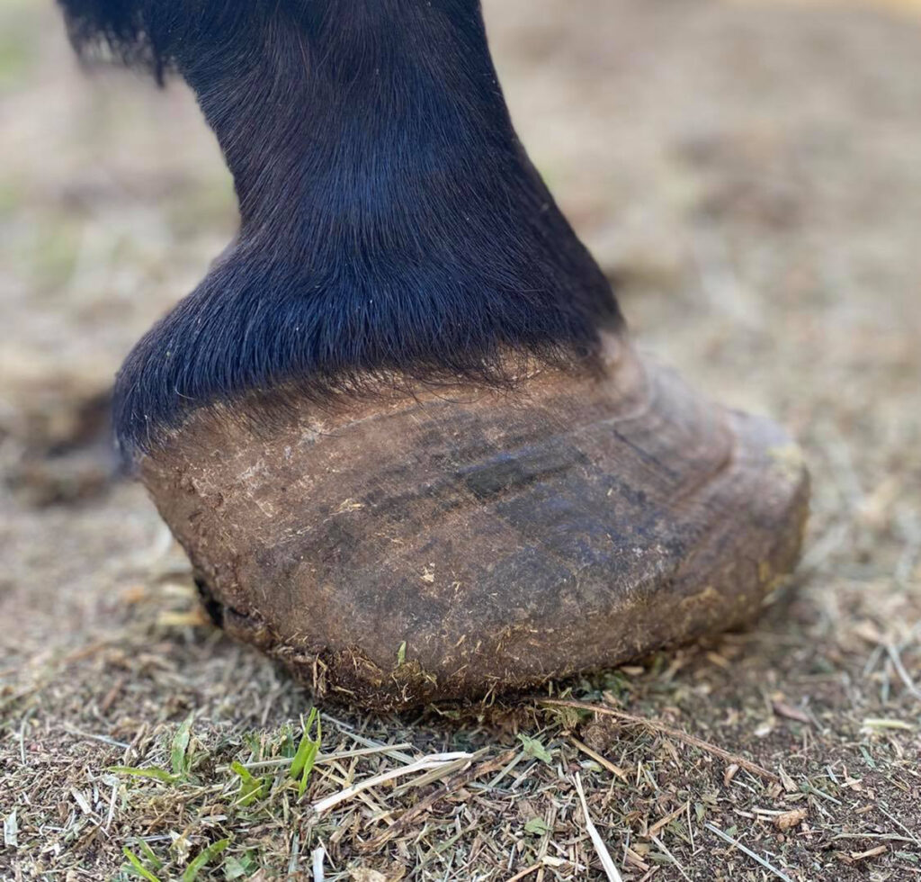
A horse that has suffered laminitis to this extent will require regular farrier attention and monitoring for the remainder of its life.
There are several illnesses that are associated with a high risk of endotoxaemia, and these include acute febrile diarrhoea, particularly Colitis and Salmonellosis; retained foetal membranes; endometritis; and pleuropneumonia. However, any bacterial disease caused by gram-negative bacteria can lead to endotoxaemia if enough endotoxin gains access to the vascular system. In cases of acute febrile diarrhoeas and pleuropneumonias, the horse is usually quite sick and undergoing intensive treatment to fight the presenting illnesses and the possibility of acute laminitis is monitored closely.
In hospital scenarios, prophylaxis treatments such as maintaining the feet in ice baths are often used to minimise the possibility of laminitis occurring. Unfortunately, in cases of retained foetal membranes, once the membranes have been removed, there can often be no indication the mare is sick or at risk of developing acute laminitis until she is found in the paddock unable to walk properly.
DIAGNOSIS NOT DIFFICULT
Diagnosing a horse with endotoxic-induced laminitis is seldom a difficult process, as the history and the clinical signs are usually very clear, particularly in cases where there is profound evidence of a severe bacterial infection followed by clinical signs associated with the feet. Initially, the horse may be seen shifting weight from one limb to the other and heat and increased pulses are detected.
As the condition begins to worsen, the horse will be seen standing with both front feet camped out in front of them and is very reluctant to move forward when prompted. Without pain relief, laminitic horses can show marked signs of stress, sweating, agitation and trembling. Some horses will lie down for extended periods to get some relief for their feet. X-raying the horse’s foot will often be performed to confirm the diagnosis.
If the veterinarian is not already involved, it is extremely important that they be consulted as soon as possible once there is any hint of laminitis occurring, as the longer the horse is allowed to progress without medical intervention, the higher the risk of permanent damage or the need for euthanasia occurring. The cornerstones of treatment are to treat (and hopefully resolve) the inciting cause of the endotoxaemia, provide pain relief, support the position of the pedal bone relative to the hoof wall, and prevent further damage to the structures within in the foot.
Treating the source of the endotoxin will depend on the cause but will often require antibiotics, anti-endotoxic medications and, potentially, intravenous fluids. Once the horse has toxic-induced laminitis, treatment will include pain relief, often using NSAIDs such as phenylbutazone, finadyne, meloxicam or firocoxib. In severe cases, stronger medications can be used to relieve pain, and these can include opioids and continuous intravenous infusions of selected pain medications.
Supporting the pedal bone can involve frog and sole supports and confining the horse to a stable with deep, soft bedding, especially if the horse is one that will lay down to relieve the weight on their feet. Otherwise, deep sand will provide some support for the feet but is not very comfortable for the horse to lay down on. If sand is used, then where possible an area of sand should be covered with straw to allow some comfort to the horse if or when it lies down.
For both affected horses and those “at risk” horses – that is, horses with illnesses that carry a higher risk of laminitis occurring – cold ice slurries are used for the horse to stand in continuously to minimise the damage to the feet. In referral equine hospitals where many endotoxic patients are admitted prior to any laminitis signs occurring, ice slurry baths are used to prevent laminitis by standing horses in these baths for 48-72 hours continuously. This is incredibly labour intensive as the ice needs to be constantly replaced to ensure the temperature of the water does not go above 10 degrees over the 2-3 days, but it has been shown to be effective.
For those horses that survive the initial few days, the inclusion of a reputable farrier with experience in dealing with laminitis is essential, as they will need to be intimately involved in the recovery and rehabilitation of the horse over the months to years that follow. EQ
