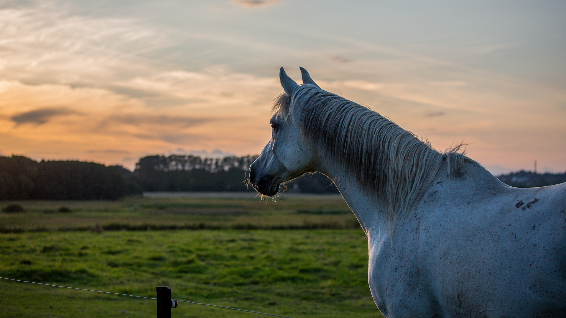

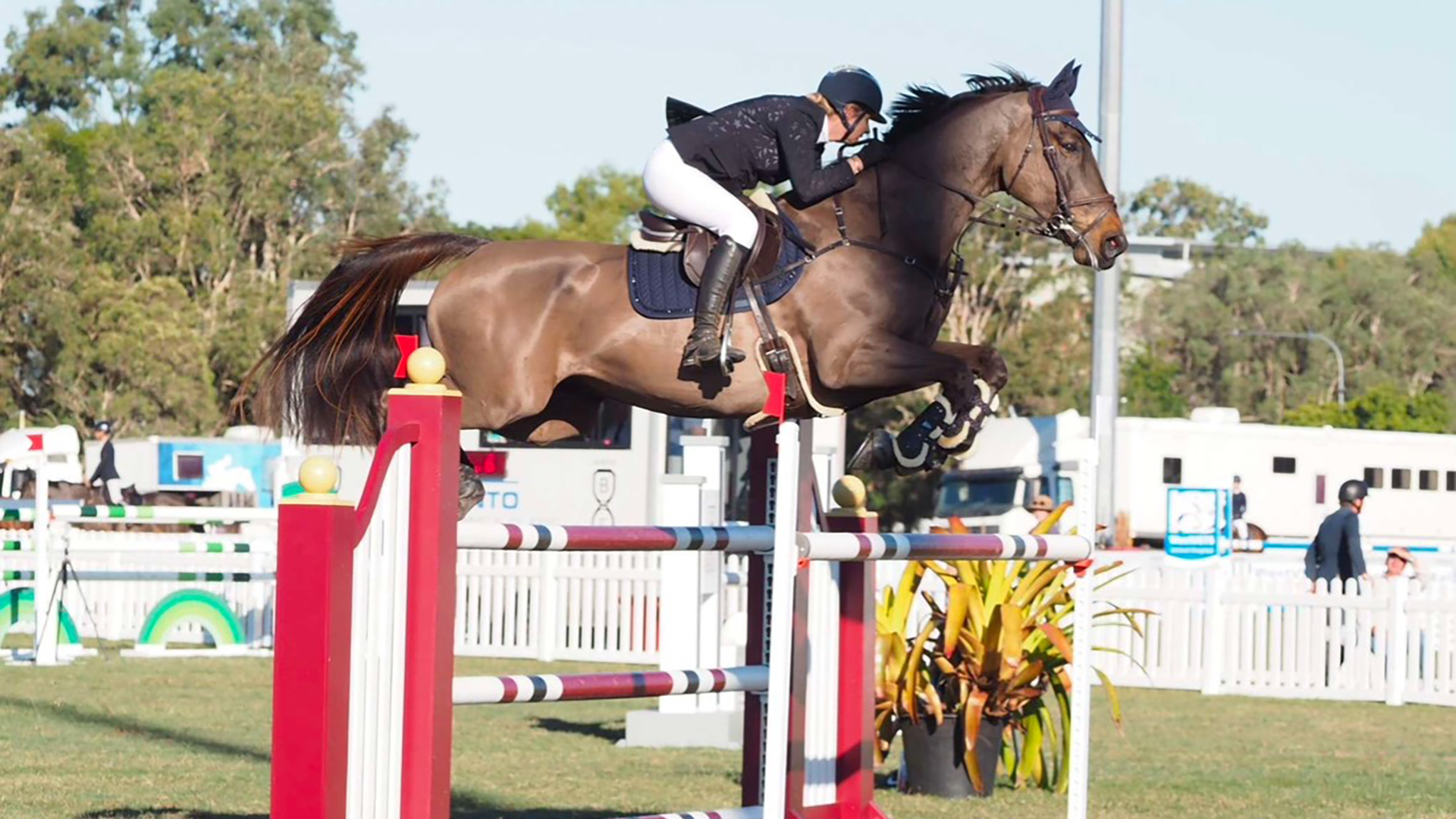
Horses, like any other mammals, can and do succumb to cancer and therefore it is important to examine our horses regularly for evidence of abnormalities that can’t be explained.
No one knows your horse as well as you do and therefore as owners you are in the best position to detect abnormalities.
This doesn’t mean you have the vet come and examine your horse every couple of months, but simply that you get to know your own horse and take the time every few months to give it a good visual look over.
Whilst skin tumours are the most recognisable tumours that we can see on our horse, discovering abnormalities such as weight loss, unexplained swellings, exercise intolerances and changes in routine behaviour can lead to early detection of cancers and potentially better treatment options and successes.
No one knows your horse as well as you do and therefore as owners you are in the best position to detect abnormalities that you can then alert your veterinarian too. Sadly, whilst the outcome for the horse diagnosed with cancer may not change, early detection does allow some cancers to be cured, some to be better managed, and some to be dealt with before the horse suffers more than it needs to.
An accurate occurrence rate of cancers in the equine world is understated as many horses die or are put down without autopsies being performed, meaning their cause of death is not recorded.
Skin cancers can usually be recorded and followed through for data collection. Melanomas are potentially overrepresented as they are often blamed for any unexplained abnormality seen in grey horses. Internal cancers are often assumed to be affecting a horse, particularly when there is weight loss in an aged individual, but they are not always accurately diagnosed. By this I mean an aged horse can become ill and slowly deteriorate, with the assumption being it has a tumour when there are other causes that could result in weight loss and death.
COMMON SKIN TUMOURS
Skin cancers can include squamous cell carcinomas (SCC), melanomas (grey horses), sarcoids, mast cell tumours, fibrosarcomas and haemangiosarcomas. By far the most common skin tumours in horses are sarcoids, melanomas and SCCs. Of these, I worry most about SCCs, as these can be very aggressive and metastasise (spread) to local lymph nodes or the lungs if not dealt with promptly.
These are commonly seen in the third eyelids and around the eyes of horse breeds that have low to no pigmentation in these areas and therefore have less protection from the sun’s UV rays compared to horses with pigmented eyelids. These tumors can invade local structures such as the eye and the bony eye socket if not removed completely. Chestnut-based colour coats, albinos and Appaloosas are more prone to develop ocular SCC than dark-coloured skin coats.

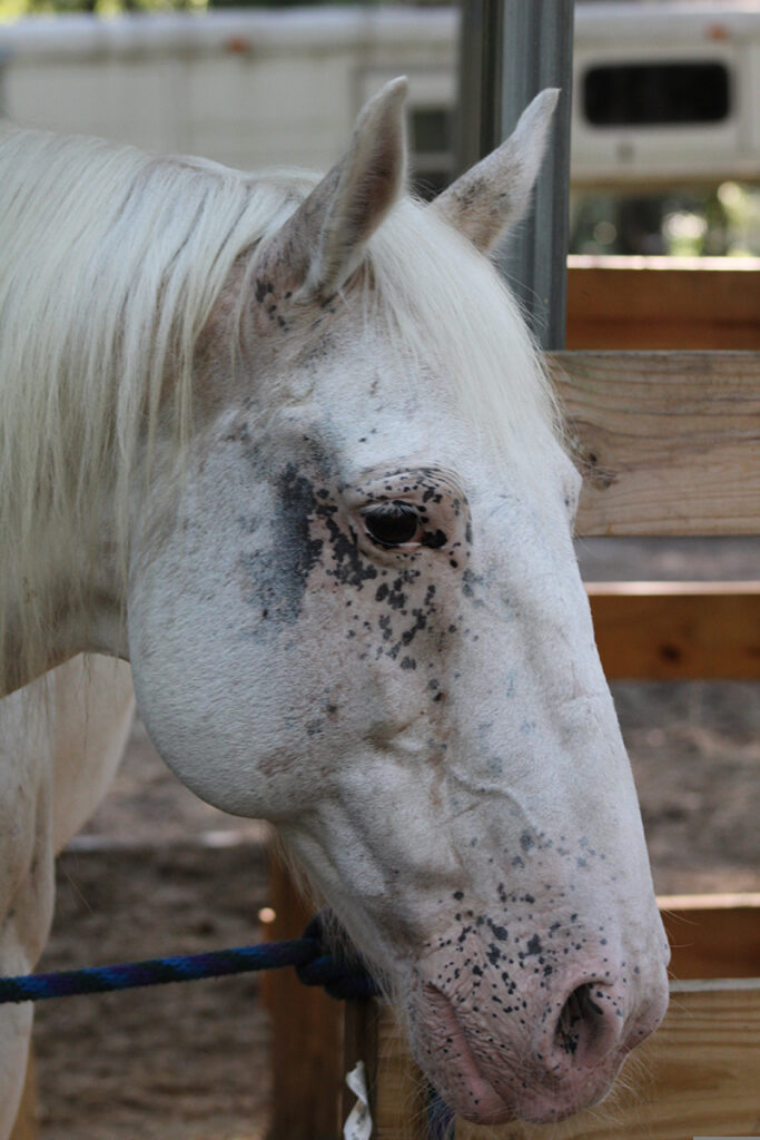
Appaloosas are a breed more prone to developing ocular SCC.
“Early detection
does allow some
cancers to be cured.”
There have also been genetic risk factors identified with certain breeds including Haflingers and Belgians that, when carrying a certain gene, greatly increases their risk of getting SCC, especially when exposed to sunlight. In Australia I would see a lot of these cancers in Clydesdales, as they commonly have large areas of their face that are non-pigmented.
SCCs are also commonly found on the penis of colts and geldings where the skin can be non-pigmented. When these cancers occur on the penis, if identified early they can be removed without the need to amputate the penis; but if left/diagnosed late, require penile amputation to save their life. They are also found on the vulval lips of mares. SCCs can also cause internal cancers that develop in the oesophagus and stomach and can metastasise to other internal structures, where they are invariably fatal.
Sarcoids are the most common skin tumours, but unlike SCCs they do not metastasise internally. They can spread locally and cause most of their damage due to their size and areas where they develop. Sarcoids have been associated with the bovine papilloma virus, however, not all horses that are infected with the papilloma virus develop a sarcoid, indicating that there are other factors such as a genetic predisposition required before a sarcoid develops.
Sarcoids come in a few different forms including verrucose (wart-like), nodular (firm round lump like), occult (flat scaley and often round areas), fibroblastic (fleshier appearance), mixed (a combination of types) and malevolent (very aggressive and fast-growing ulcerative nodules). These tumours can be difficult to deal with as sometimes they sit dormant without changing/growing for years. But other times, once aggravated, they can grow and spread rapidly over a large area.
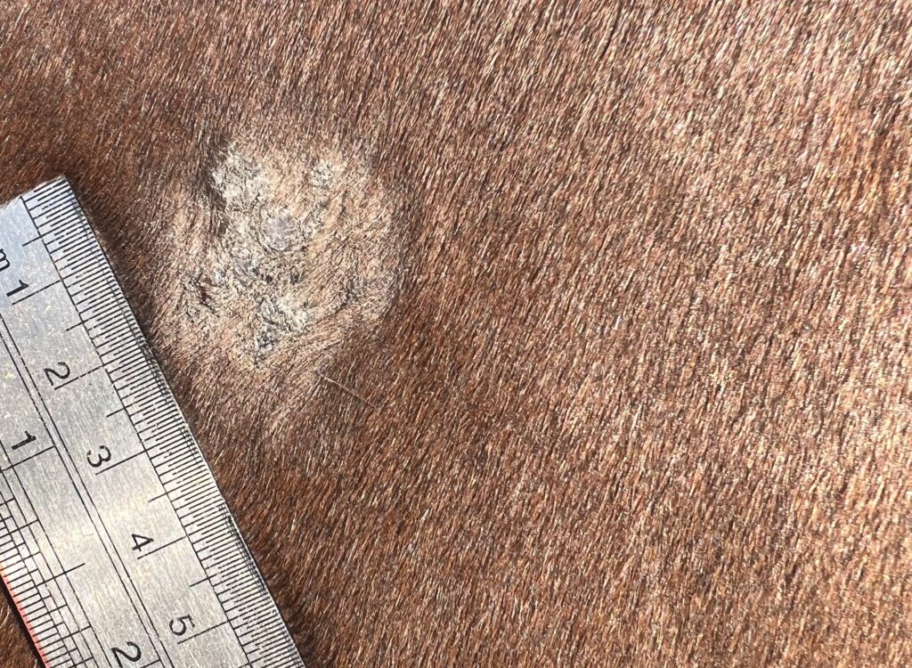
A sarcoid; you can monitor skin tumours with a ruler to ascertain how quickly they growing. Image by Dr Maxine Brain.
TREATMENT THE SOONER THE BETTER
There are multiple treatments available depending on the type, size and position of the sarcoid. Generally, the smaller and the more quickly these tumours are treated/removed, the better the long-term prognosis for the horse. If located in areas where the tack is going to rub them, they are best removed, as the constant irritation will stimulate their growth. For large sarcoids or those in areas where surgical removal is not possible, other treatments such as cryotherapy and topical chemotherapy are used.
“If located in areas where the
tack is going to rub them,
they are best removed…”
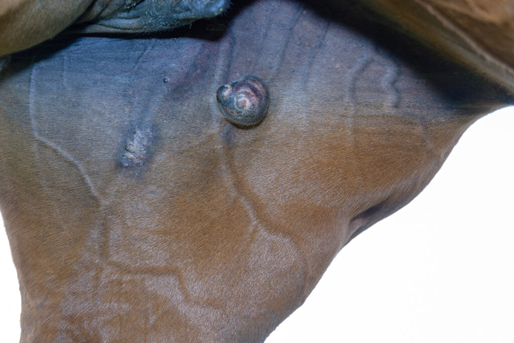
A sarcoid on the inside of the hind leg.
Melanomas are commonly found in the skin of grey horses but their classification as cancers is debatable. Most grey horses will develop skin melanomas in their lives, commonly around the anus, vulva and the butt of the tail, but they can also be found commonly around the eyes, mouth and behind the jowls, in what is known as the parotid region. They appear as nodules and contain deposits of melanin, a black pigmented substance and are easily diagnosed because of this pigment.
Many of these melanomas are benign (“not cancerous”) but grow very large or are present in such large numbers that they can interfere with defecation (passing manure) or mating. Some melanomas will transform into malignancies (cancer) that grow rapidly and metastasise internally. Melanomas that develop internally or are located internally as a metastasis will present with clinical signs associated with the organ they are affecting; for example, a melanoma can cause colic if affecting the gastrointestinal tract.
The number of sites they have been detected is numerous. Melanomas can develop in the spinal cord, meaning as they grow they put pressure on the spinal cord, causing neurological signs such as ataxia or incoordination. They can grow in the eye and cause blindness, and they can often be found in the guttural pouches, which are pouches associated with the throat that contain some large nerves and blood vessels, with tumours here leading to bleeding and neurological abnormalities.
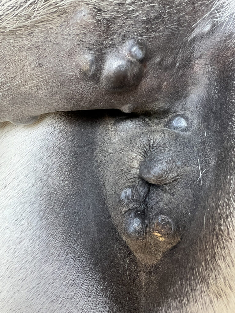
Most grey horses will develop skin melanomas in their lives, commonly around the anus, vulva and the butt of the tail. Image by Dr Maxine Brain.
Melanomas can also occur in non-grey horses, and in these cases are far more likely to be malignant. So, any non-grey horse that has a tumour that looks like a melanoma (black), should be monitored far more closely and removed, if possible, as these tend to be more aggressive.
Other skin tumours such as mast cell tumours, cutaneous lymphomas and fibrosarcoma are less common but can be differentiated using a biopsy and removed if present in a site amenable to surgery.
INNUMERABLE INTERNAL TUMOURS
Listing all internal cancers would be exhaustive for this article but suffice to say that there are multiple types of internal tumours that can affect the horse, and sadly many of them will result in the death of the horse. These include lymphosarcoma, ovarian tumours, testicular tumours, mammary gland tumours, liver tumours (cholangiocarcinomas, hepatocellular), gastric tumours, lipomas and leiomyosarcomas (smooth muscle tumours).
Lymphosarcoma deserves a special mention as it can occur in young horses, whereas most of the other cancers are seen more in middle-aged to elderly horses. Lymphosarcomas can occur in young horses and I have seen several in two and three-year-old horses, which is always a sad scenario.
Typically, these young horses present with weight loss despite a voracious appetite. Owners stress about ensuring their horse has been wormed and had its teeth done, but despite their doing everything they can to put weight on the horse, the horse continues to eat and eat but loses weight dramatically, quickly becoming a walking skeleton. These tumours are usually identified in the lymph nodes of the horse but seldom can be cured. Cortisone can be used to modify the symptoms; however, a prompt diagnosis and euthanasia is usually the best option.
Mares can get granulosa cell tumours on one or both ovaries and these are normally detected after the mare displays clinical symptoms related to hormonal changes brought about by the tumor cells. They can be provisionally diagnosed using a rectal ultrasound but often require confirmation using blood tests. Some of these tumours can become very large before they are detected; with some I have diagnosed the size of a basketball.
Whilst most are suspected because of the behavioural changes, there have been a few that have been detected due to other clinical symptoms such as abdominal pain when working (imagine a basketball-size growth swinging in the abdomen during exercise) and refusal to jump or perform. Retained testicles in male horses are susceptible to tumour development and therefore this should be considered in cryptorchid horses that develop vague symptoms of ill health and weight loss.
Diagnosing many of these internal cancers generally relies on blood tests to identify which organ is affected, ultrasonography to visualise the size and position and a biopsy to accurately determine the type of cancer if the tumour is accessible from the outside. In some cases, taking a sample of fluid from the abdomen or the chest may be helpful in diagnosis as the fluid might contain cancer cells that have migrated from the tumour.
Stomach cancers can be diagnosed using gastroscopy, but it is difficult to see much further than the upper section of the duodenum (first part of the small intestine).
In some situations where the horse is valuable or the owners are happy to do everything possible, an exploratory abdominal surgery is performed to gain conclusive proof that the horse has cancer. In rare cases, where the tumour is solitary and easily accessible, the growth can be removed but if the tumour has metastasised, euthanasia is usually performed on humane grounds.
IMPROVED SUCCESS RATES
Options available to treat skin cancers have improved immensely with surgical removal, topical chemotherapies, electrochemotherapy and immune modulators becoming more readily available and therefore improving the success rates that can be achieved.
Sadly however, the treatment options for internal cancers are not as favourable as treating skin cancers with chemotherapy being costly due to the size of the horse. Added to this is the fact that many cancers are not diagnosed at an early enough stage to be effective enough for removal prior to them spreading throughout the body or advancing to a stage where surgical removal is not practical.
There are, however, research studies occurring overseas that are showing good results with chemotherapy without the horse suffering the terrible side effects we associate with chemotherapy in humans. Studies show affected horses can go through treatment without compromising their quality of life too much. Whilst readily available in some countries, affordable chemotherapy is not currently available here, though advances in the future may see this improve. The key to successful treatment will always be an early diagnosis, so take the time to know your horse and seek veterinary help when you detect a problem. EQ
Related articles
A lacklustre coat, lack of energy, loss of muscle tone and weakened immune system were the vague yet worrying symptoms displayed by top show jumper ‘Alan’. Rider Gemma Creighton knew something wasn’t right; however, it wasn’t until a friend mentioned myofibrillar myopathy that the penny dropped.
Sedation is a common procedure in the equine world and as vets, we would be lost without having safe, reliable and easily administered drugs to use. We use sedation to quiet the horse, which allows us to perform a huge variety of procedures, reducing the risk to horse and human and allowing minimal interference so a procedure can be performed quickly and efficiently.