Much like the extreme discomfort humans suffer when choking, it is equally distressing when a horse chokes – and the treatment is much more involved than judicious thumps between the shoulder blades.

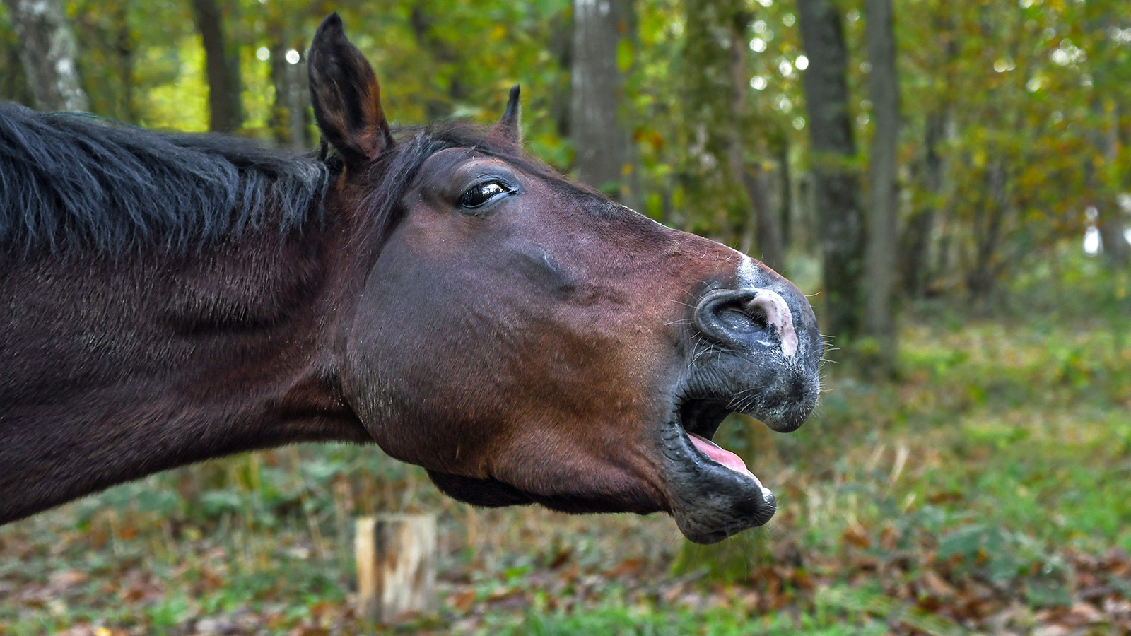
When choke occurs in horses, food and saliva comes out of the nostrils due to obstruction of the oesophagus. Image by Dr Maxine Brain.
Saturday night, and what should have been a quiet night after a long day out competing, is quickly turned into a night of concern and panic. Timmy the six-year-old thoroughbred gelding appeared to be fine coming off the float, but five minutes after being let go in his box, he is making retching motions with his neck and has green stuff coming from his nostrils. The longer Timmy is left, the more distressed he becomes, leaving his owners stressed and trying desperately to contact a veterinarian.
What has happened to Timmy is not an uncommon scenario, but it is a frightening one. A careful history taken over the phone and I am reasonably confident that Timmy has choke and instruct the owners not to give him any food or water, and I drive straight there. Whilst not a life-threatening situation in the short-term, it is hard to leave a horse visibly distressed any longer than necessary.
Choke in a horse is different to what we would expect with a person choking. A horse that is choking has food or a foreign body lodged in its oesophagus, the part of the gastrointestinal tract (GIT) that is responsible for moving food from the pharynx (throat) to the stomach. In people, choking refers to the airways becoming blocked, and the patient is unable to move oxygen into their lungs. When a horse has choke, it can breathe but nothing can pass from the pharynx into the stomach, including the saliva that is produced, resulting in the buildup of fluid that has nowhere to go other than back up through the nasal passages and out of the nose.
Horses can choke on many things; for Timmy it was one of the commoner causes, hay. Coming home after a hard day of competition, horses can often be a little dehydrated and hungry. The horse takes a big mouthful of hay, but doesn’t chew the food properly or release enough saliva to mix with it sufficiently to help lubricate the hay. Once they swallow the bolus of semi-lubricated hay, it becomes lodged at some point between the pharynx and the stomach.
Although the bolus can become stuck anywhere, it is more common for it to lodge in one of three areas along the tract where there is an anatomical or mechanical reduction in the ability of the oesophagus to dilate and let the bolus move through. The common sites are the mid cervical region (middle of the neck), the thoracic inlet (where the oesophagus moves into the chest) and the last part of the oesophagus just before the sphincter that opens into the stomach.
When the hay is lodged in the mid cervical region, it can often be seen or palpated, although this can be painful for the horse and cause it to tense its neck. When lodged at the thoracic inlet or near the stomach, it can’t usually be seen or palpated, and the location is estimated by how far a stomach tube can be passed before a blockage is met. Timmy had a blockage around the thoracic inlet, and I was unable to pass a stomach tube down very far before its passage was stopped.
“Choke in a horse is different to what
we would expect with a person choking.”
BENEFITS OF SEDATION
The first thing I normally do when faced with a choking horse is to sedate it and there are two reasons for this. The first is it relaxes the horse and the muscles within the oesophagus, making it more comfortable to pass a stomach tube. As was the case with Timmy, horses can get very distressed and anxious, sometimes trying to lie down to relieve the discomfort, so giving them sedation helps reduce the stress and the pain. The second benefit of sedation is it encourages the horse to stand with his head lowered so any saliva or feed that banks up in the oesophagus can run down the nasal passages and out through the nostrils onto the ground. Sometimes, the sedation will relax the horse sufficiently to allow the blockage to be moved into the stomach without any further intervention. This unfortunately wasn’t the case for Timmy.

Many things can lead to choke in horses, but hay is one of the most common causes.
“Horses can choke
on many things.”
Treatment is generally centred around passing a stomach tube – and either gently moving the obstruction along the oesophagus and into the stomach – or passing a tube and slowly lavaging or flushing water through the tube into the oesophagus. Lavaging can help break up the feed and let some come back out through the nose, often reducing the mass sufficiently that it can be moved on and through the oesophagus.
Fortunately, this was all that was required with Timmy. Although it took 20-30 minutes, a lot of gentle manoeuvring with the tube, and multiple administrations of water to flush out pieces of semi-chewed hay, I was able to unchoke Timmy relatively easily. Unfortunately, this has not always been the case with other chokes. It is important to note here that mineral oils or lubrication should never be used to help flush a choke, as the risk of them being aspirated back into the lungs is high, and the consequences can be catastrophic.
The most severe cases of choke I have encountered have involved rigid objects such as wood (from eating a tree), apples and pieces of carrot. These products are unable to be lavaged free or broken up with water. It takes a lot of time, patience, and sedation to relieve these types of obstructions and I have spent several hours on multiple occasions trying to resolve choke. It is exceedingly important that any manipulation done with the tube is gently performed as there is a big risk of perforating the oesophagus wall if the person doing the tubing is too rough.
IDENTIFYING THE OBSTRUCTION
For some cases of choke, I have used an endoscope passed down the oesophagus to observe what is stuck when the owner is unsure of what the horse has eaten. Knowing what the horse is choking on can influence how I proceed. For obstructions like hay or fibrous materials I have passed biopsy forceps through the channel of the scope to grab small pieces of wood or hay to help debulk the trapped mass. This can be very satisfying to watch when you pull out some of the mass and suddenly see the rest disappear down the oesophagus as a wave of muscular contraction takes over and moves the blockage through. The easiest blockages to remove are usually those caused by pellets or grain feed as these can be flushed out in small amounts with the tube and water and, in my experience, the most likely blockages to resolve on their own or with sedation alone.
There have been cases where the obstruction has not resolved with the traditional treatments and a different tactic is used. One of these is giving the horse a drug called oxytocin, as this causes smooth muscles to contract very strongly. The idea behind this is the muscles contract so hard, the obstruction is forced down. I have seen this used occasionally but the owner should be advised that there is a risk that too strong a muscular contraction on a rigid, firmly lodged foreign body does have an increased risk of damage/perforation of the oesophageal wall.
Surgery has also been tried for cases that are refractory to every other medical attempt, however, the risk of wound dehiscence (breakdown) and scar formation leading to ongoing problems with swallowing have made this less appealing and more a last resort.
Whilst Timmy was a straightforward case with little risk of ongoing complications, there are several things that need to be assessed and monitored in the days to weeks that follow. One of the big risks with choke is feed, saliva or water coming up from the oesophagus that can be aspirated into the lungs and cause pneumonia. This is why it is important to withhold all food and water when the horse is first noted to choke. As stated above, sedating the horse and encouraging it to stand with its head down reduces the chances of the feed going into the lungs, as the pharynx is lowered below the chest and anything in the pharynx will run down the nose and not up the trachea. When a horse holds its head up, the pharynx is above the chest and any feed or water that escapes into the trachea will run down into the lungs and cause an infection, that progresses to pneumonia.
Another complication, particularly with chokes that have been ongoing for hours to days, is significant damage to the oesophagus due to bruising or tearing of the mucosa and muscle where the obstruction has placed sustained pressure to one area. If the damage is superficial, it can heal without issue, but with deeper injuries there is risk that the oesophagus will heal with scar tissue. This is undesirable as scar tissue has no stretch properties, increasing the risk of further episodes of choke occurring.
When large areas are damaged, strictures or tight bands of tissue can form and these lead to more frequent episodes that can perpetuate the damage. In severe cases, veterinary intervention is required to try and gradually stretch the scar tissue, using specialised equipment called bougies or dilation balloons. Horses with known strictures need to be managed to minimise chokes occurring and this can include slowing their eating, and feeding only chaffed or pelleted feeds. Moistening feed can also be beneficial in some cases.

It can be risky feeding horses hay in the float on the way home from an event if they are tired and dehydrated.
MANAGEMENT PROTOCOLS
This was the first episode of choke for Timmy so, after several days of monitoring his temperature and observing him eating with no recurrences, he was given the all-clear. For horses that suffer recurrent bouts of choke, special management protocols should be put in place to reduce the risk of this occurring. Management practices will be determined based on the reason the horse has obstructed. For Timmy, it was having access to hay when dehydrated and exhausted; owners would be advised that the next time he has a big competition, allow him to go to his box, have some water and settle before feeding him, preferably bucket feed and avoid hay until the horse is fully recovered. It can be risky feeding horses hay in the float on the way home from an event for this same reason.
Horses that choke frequently but seldom leave home or exercise can have other underlying issues that predispose them. Older horses that choke, especially on hay or carrots, should have their teeth checked by the dentist. Razor-sharp points and missing teeth can reduce the amount of chewing a horse does and result in them swallowing a larger bolus of food without thorough mastication. Younger horses can also have dental abnormalities that predispose them to choke, but this is less common.
Younger horses that gulp their feed can have the same scenario whereby they don’t masticate the food properly and swallow large, under-lubricated feed. This is seen in paddocks where there is competition for feed and one horse rushes in and takes a big mouthful before his paddock mates chase him off, or simply gutses the food out of fear it will miss out. This can happen when feeding time has been delayed for several hours and the horse thinks he is starving. In this situation there are several management options, including feeding horses prone to choke in a yard by themselves, placing large bricks in their feed bins so they need to forage around the brick and can’t take big mouthfuls, or feeding smaller amounts initially if food has been withheld for hours for other reasons.
There is also the rare case of oral or oesophageal tumors that can cause partial obstruction of the GIT and predispose the horse to choke because of a narrowed lumen (opening). These would be rare in my experience and sadly would be more likely to have a poor prognosis, as many of these types of tumors would not be amenable to surgery. EQ
YOU MIGHT ALSO LIKE TO READ BY DR MAXINE BRAIN:
The Challenge of Treating HPSD – Equestrian Life, May 2023
From the Horse’s Mouth: Salivary Glands – Equestrian Life, February 2023
Cardiac Murmurs – Equestrian Life, February 2023
Matters of the Heart – Equestrian Life, January 2023
Umbilical Concerns in Foals – Equestrian Life, December 2022
Retained Foetal Membranes – Equestrian Life, October 2022
Preparing for Laminitis – Equestrian Life, September 2022
Working Together for Best Outcomes – Equestrian Life, August 2022
What Constitutes an Emergency – Equestrian Life, July 2022
Peri-Tarsal Cellulitis Calls for Quick Action – Equestrian Life, June 2022
Sinusitis: Not To Be Sneezed At – Equestrian Life, May 2022
Japanese Encephalitis: No Cause For Alarm – Equestrian Life, April 2022
Hernia Learning Curve – Equestrian Life, March 2022
Osteochondromas: Benign But Irritating – Equestrian Life, February 2022
Don’t Forget the Water – Equestrian Life, January 2022
Understanding Anaesthesia – Equestrian Life, December 2021
A Quick Guide to Castration – Equestrian Life, November 2021
Caring for Mammary Glands – Equestrian Life, October 2021
Sepsis In Foals – Equestrian Life, September 2021
Understanding Tendon Sheath Inflammation – Equestrian Life, August 2021
The Mystery of Equine Shivers – Equestrian Life, July 2021
Heads up for the Big Chill – Equestrian Life, June 2021
The Ridden Horse Pain Ethogram – Equestrian Life, May 2021
The Benefits of Genetic Testing – Equestrian Life, April 2021
Heavy Metal Toxicities – Equestrian Life, March 2021
Euthanasia, the Toughest Decision – Equestrian Life, February 2021
How to Beat Heat Stress – Equestrian Life, January 2021
Medicinal Cannabis for Horses – Equestrian Life, December 2020
Foal Diarrhoea Part 2: Infectious Diarrhoea – Equestrian Life, November 2020
Foal Diarrhoea (Don’t Panic!) – Equestrian Life, October 2020
Urticaria Calls For Detective Work – Equestrian Life, September 2020
Winter’s Scourge, The Foot Abscess – Equestrian Life, August 2020
Core Strengthening & Balance Exercises – Equestrian Life, July 2020
The Principles of Rehabilitation – Equestrian Life, June 2020
When is Old, Too Old? – Equestrian Life, May 2020


