This article has appeared previously with Equestrian Life. To see what’s in our latest digtal issue, click here.
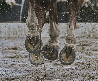
No hoof, no horse!
It has been estimated that as much as 70% of all the lameness problems in performance horses are hoof-related. Many, if not most, of the problems could be avoided if the time is taken to evaluate the horse’s feet properly and take proper precautions.
When you are evaluating your horse’s feet, several things should be considered. First, examine the hoof wall for cracks. A brittle wall is a potential problem because it may let in dirt, bacteria or fungus, which can lead to abscesses, seedy toe, gravel and white-line disease. All of these conditions may result in a lame horse, something we would like to avoid! A brittle, cracked wall maybe a genetic problem. A bad-footed mare often produces a bad-footed foal. If your horse has a problem, putting shoes on can help because it holds the foot together and less chipping will occur. Adding biotin and methionine to the diet will also help produce a better, thicker wall. These additives are easy to find, are cheap, and they really do help.
Evaluate the shape of the foot. Is there too much toe? Is the heel angle too far under the foot, indicating an under-run heel? Is the sole of the foot too close to the ground, indicating flat footedness? These are all important questions to answer if one is to fully evaluate the foot’s shape. Again, shape abnormalities predispose the horse to lameness. Corrective shoeing can help minimise these problems; a good farrier is critical.
Hoof condition is normally a reflection of the quality of nutrition that a horse is receiving. Each horse is an individual and has its own special requirements of minerals and vitamins. Some horses digest food better than others and so need a different proportion of minerals and other nutrients in the diet. Some horses only produce thin hoof wall and sole and the rate of growth is slower than normal, often causing hoof problems.
Horses often become uneven moving from soft winter grounds to dry summer surfaces or visa versa. What has happened here is that your horse’s hooves are often not as strong as they could have been right from the previous season but could cope whilst the ground was soft or hard. Only when the ground changes do they start to feel their feet. The answer is to supplement their diet within the prior season, not to wait until they go lame.
Hoof is a relatively slow growing tissue, it takes about nine months for a hoof to grow down from coronary band to the ground, so you need to ensure that the horn is as strong as possible before his feet are subject to hard or rough going. Many horses with poor feet get by on clay, but when changed to a sandy soil may go lame. This is because the sand fills up the bottom of the foot and adds sole pressure. Thin-soled horses or those with flat feet have trouble. Clay soil also holds moisture better than sand and therefore adds more moisture to the foot. A brittle hoof wall will get worse as it dries out. Hence, in the sandy soils good foot care takes on a whole new meaning.
HOOF CRACKS These can be superficial defects or severe problems that cause lameness. They can stem from a variety of causes, including laminitis, trauma, repetitive concussion on a hard surface, or unbalanced feet. Vertical hoof cracks are common in horses with poor hoof care or those with excessively dry, shelly hooves. An injury to the coronary band can also result in a vertical hoof crack. Internal imbalance can cause surface hoof cracks in even the best of shoeing.
Hoof cracks provide an opportunity for pathogens to invade the hoof wall, which can lead to lameness-causing infections and white line disease. Some hoof cracks require external fixation. Screws and surgical steel wire are used to lace the crack together to prevent additional damage.
Sandcracks can be treated with nail or toe clips either side of the fissure. This will stabilise the area enough for supplements to do their work in growing new horn. If the crack is growing up to the coronary band it needs to be stopped before it damages this area and causes permanent damage. In such cases a bar is inserted at the top of the crack to stop it spreading further. When a horse has deep sandcracks they may need to be sewn together with steel wire and crack filled in with resin.
THRUSH This is a bacterial infection of the horse’s frog and sole. It is prevalent in horses that are kept in unclean conditions such as damp stables or paddocks, but can occur in any horse. Thrush-infected feet produce an offensive odour and a black discharge around the frog. If thrush is allowed to progress far enough so that the sensitive areas of the hoof are affected, lameness will result. Several treatments with an iodine or phenol product usually kill the pathogens involved. To prevent thrush from occurring or to help a horse recover from it, owners should perform proper hoof hygiene (regularly pick debris from the hooves), maintain clean, dry stall/pasture footing, and schedule routine farrier visits.
SEEDY TOE occurs when layers of the inside of the hoof wall separate at the white line, creating a cavity which starts from the sole and may extend right up to the coronet. The layers that separate are the sensitive and insensitive laminae layers. The cavity of seedy toe can be seen on the underside of the foot as a soft area around the white line. It is rather like a fungal infection. This is where the cavity is being filled in with crumbling weak horn.
When seedy toe causes problems, dirt manages to get into the cavity and create infection. There are differences of opinion as to why seedy toe starts but it is likely to be due to either damage done by laminitis or by poor nutrition resulting in weak horn growth. Poor nutrition can also be a result of inadequate digestion of good food eaten.
Treatment: Cavities can be cleaned thoroughly and filled with cotton wool, Stockholm tar or other commercial products for this purpose. A broad webbed shoe will hold the dressing in place. If the cavity cannot be filled easily the overlying horn can be taken away in order to allow fresh healthy horn to grow down. If infection occurs a foot will need to be tubbed and poulticed regularly. After treatment supplements are necessary to improve horn growth and to help prevent recurrence of laminitis.
ABSCESSES can cause sudden and sometimes severe lameness in horses. These are localised accumulations of pus between the germinal (growth layer) and keratinised layers of the hoof wall. They are found most commonly in the sole, but can be located elsewhere within the hoof. The infection and pressure of pus accumulation can cause severe pain. In principle the laminae (the most sensitive part of the hoof) is inflamed and infected due to various injuries. These can be caused by pricking the horse’s foot with a sharp object such as nails, various bruises due to an impact that can get infected in a latter stage as seen often in the winter months on bare footed horses. Excessive pressure to the laminae (usually under the bars) will cause tissue to die off and in time will cause an abscess. Foreign objects (stones, dirt, bacteria) working their way through the horny part of the sole or along side/beneath the wall to the sensitive part of the hoof (laminae) and in some cases thrush can also cause an abscess of the laminae above the frog.
Hoof abscesses can be caused by sole penetration by a sharp object (introducing pathogens deep within the hoof), bacterial invasion of a hoof crack, or infection of a bruised sole. ‘Gravel’ is sometimes used as a lay term to describe abscesses that are forced up along the hoof wall to break and drain at the coronary band.
Early Stages: Typical signs of hoof abscesses include acute to severe lameness where the horse bears minimal to no weight on the affected limb, an increased digital pulse, and heat in the limb. Hoof abscesses are typically superficial and always affect the sensitive tissue of the hoof. Leaving abscesses untreated can undermine the foot’s integrity, leading to a prolonged and often costly layup period.
Treatment: ‘Treatment for superficial abscesses usually means thoroughly cleaning the hoof and locating the point of entry or the road map that says this is where the abscess is,’ Redden says. ‘Then, opening a very, very small hole to allow drainage (about the size of a pencil tip). The horse will usually exhibit instant relief when the pressure built up by the trapped infection is released. A very large hole will cause prolapse of the sensitive tissue and create a larger problem than you had initially.’
It’s important to protect the area while the hole is allowed to heal. Bandage the foot with a betadine (never use a strong iodine solution) wrap, and use a hospital shoe or pad to help protect it.
WHITE LINE DISEASE (WLD) is a term used to describe a keratolytic process (one that destroys the structural protein keratin) on the surface of the hoof, which is characterised by a separation of the inner zone of the hoof wall (which is white in colour). It is the separation of the inner hoof wall from its outer layers caused by opportunistic pathogens. This separation occurs within the stratum medium. The process is usually secondary to a mechanical hoof problem that causes an initial hoof wall separation.
These problems might include flexural deformities (such as contracted tendons), club foot, chronic laminitis, hoof cracks, chronic infection (abscesses or gravel), injuries that cause scarring of the stratum medium, chemical imbalances, or direct trauma with subsequent bleeding.
Early stages: WLD is not easily detected. It is characterised by a small, powdery area located in the white zone of the hoof wall. As the horn wall deteriorates, the sole pushes forward and covers the deterioration as fast as it occurs.
When a farrier pulls the shoe off, he can’t see that white chalky area. X-ray is the only way to detect these early changes. The disease can be isolated in one foot, or it can affect multiple feet. It is most often found in a club foot or a foot with high heels. The disease actually begins when the fungus and bacteria invade a weakened area of the horn wall or a pre-existing scar. WLD most commonly affects the toe and the interior quarters of the foot. Pathogens invade these openings and progressively destroy the inner hoof wall. Factors contributing to this situation are not well defined.
Treatment: Varies according to hoof involvement and the severity of the disease. Applying a rocker shoe to reduce the DDFT (deep digital flexor tendon) tension can work very well in the majority of cases without having to remove a large area of the hoof wall.
However, when damage to the hoof is too severe, removing the infected areas of the hoof wall (resection) is necessary to allow access to treat the pathogens. Once the veterinarian or farrier removes the infected areas, he or she soaks the foot in a chlorine-based solvent to help kill any pathogens that are still present. Depending on the degree of infection, the hoof wall can take several weeks to several months to heal. Even horses kept in the most immaculate environment can be affected with white line disease as it is not directly related to hygiene or negligent farrier care.
Every acute foot problem has the potential to be an emergency. Therefore, never ignore the lameness and hope that it will go away. Call your farrier and veterinarian for a consultation right away. Together, they can quickly determine the significance of the problem and establish a course of treatment.
READ THE LATEST NEWS ARTICLES HERE


